
Page 69 - 14_Atti_SIN_SNO_2022_flip
P. 69
Proceedings SNO “Percorsi clinici in Neuroscienze”
A B C D E
F G
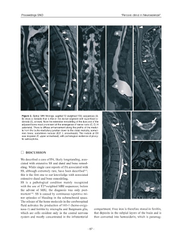
Figure 2. Spinal MRI findings: sagittal T2 weighted TSE sequences (A,
B) show a medulla that is thin in the dorsal segment with superficial si-
derosis (G, arrows). Note the extensive remodeling of the dura and of the
adjacent bone most prominent at the emergence of nerve roots (C, D, H
asterisks). There is diffuse enhancement along the profile of the medul-
la from the bulbo-medullary junction down to the distal medulla, someti-
mes linear, sometimes nodular (E,F, I, arrowheads). The nodule at D3
was biopsied (F, upper arrowhead), with pathological evidence of pilocy-
tic astrocytoma.
H
DISCUSSION
We described a case of PA, likely longstanding, asso-
ciated with extensive SS and dural and bone remod-
eling. While single case reports of PA associated with
SS, although extremely rare, have been described ,
(6,7)
I
this is the first one to our knowledge with associated
extensive dural and bone remodeling.
SS is a pathological condition mainly recognized
with the use of T2*weighted MRI sequences; before
the advent of MRI, the diagnosis was only post-
(7,8)
mortem . SS is caused by continuous repetitive mi-
nor episodes of bleeding in the subarachnoid space.
The release of the heme molecule in the cerebrospinal
fluid activates the production of HO-1 (hemo-oxige-
nase-1) and ferritin by microglia and Bergmann glia, compartment. Free iron is therefore stored in ferritin,
which are cells resident only in the central nervous that deposits in the subpial layers of the brain and is
system and mostly concentrated in the infratentorial then converted into hemosiderin, which is paramag-
- 67 -